Skin cancer is the most common form of cancer worldwide, according to the Skin Cancer Foundation Australia. Early detection is critical; if melanoma is caught early, the survival rate is extremely high. To improve diagnostic accuracy, dermatologists rely on a specialised optical instrument called a dermatoscope, which allows clinicians to examine skin lesions in far greater detail than the naked eye can reveal.
Why Early Detection Matters
Skin cancer presents in several forms, including melanoma, basal cell carcinoma, and squamous cell carcinoma. Cancer Council Australia recommends routine skin examinations because suspicious lesions can develop in areas that are difficult for patients to monitor themselves.
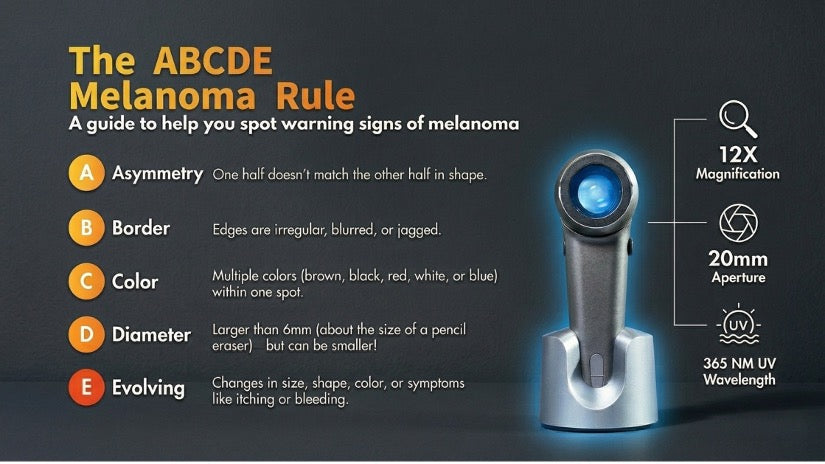
Dermatologists are trained to identify warning signs using the ABCDE rule of melanoma detection, a guideline promoted by the National Cancer Institute:
-
Asymmetry in moles
-
Irregular borders
-
Uneven colouration
-
Rapid changes in size or shape
However, many early skin cancers display subtle structures beneath the skin surface that cannot be seen without magnification and specialised lighting, and this is where dermoscopy becomes essential.
What Is a Dermatoscope?
A dermatoscope is a medical imaging device used by dermatologists and other healthcare professionals to examine skin lesions with enhanced clarity. It combines three essential elements:
-
Magnification to enlarge skin structures
-
Polarised or non-polarised lighting to illuminate deeper layers of the skin
-
Optical lenses
Together, these features allow clinicians to visualise pigment networks, vascular patterns, and keratin structures that remain invisible during a standard visual exam. This diagnostic technique is known as dermoscopy, or dermatoscopy.
How Dermoscopy Improves Diagnostic Accuracy
Multiple clinical studies demonstrate that dermoscopy significantly improves the accuracy of skin cancer detection. Research published on the National Library of Medicine indicates that dermoscopy can improve melanoma detection rates by up to 90% compared compared to 81% with visual examination alone
Key benefits include:
-
Earlier identification of suspicious lesions
-
Reduced unnecessary biopsies
-
Improved monitoring of evolving moles
-
Better differentiation between benign and malignant lesions
Because of these advantages, dermoscopy has become a standard tool in modern dermatology practice.
What Dermatologists Examine During a Dermoscopic Assessment
When using a dermatoscope, clinicians evaluate specific visual patterns within the skin. These microscopic features are only visible when magnification and proper lighting are applied:
-
Pigment networks: Mesh-like patterns that help differentiate benign moles from melanoma
-
Vascular structures: Blood vessel patterns that can reveal inflammatory skin diseases or malignancies
-
Keratin structures: Certain formations that can indicate non-melanoma skin cancers such as basal cell carcinoma
The Importance of High-Quality Instruments
The clarity and accuracy of a dermoscopic exam depend heavily on the quality of the optical instrument being used. Modern dermatoscopes provide higher magnification levels, improved illumination systems, enhanced image clarity, and better portability for clinical environments.
One example is the ILLUCO IDS‑9100 Dermatoscope, which supports detailed skin examinations with 12× optical magnification and precision optics.
Key features include:
-
High-resolution optical magnification
-
Clear visualisation of skin structures
-
Durable construction for clinical use
-
Ergonomic design for comfortable examinations
When to See a Dermatologist
Patients should seek evaluation if they notice new or changing moles, lesions that bleed or do not heal, rapidly growing spots, or irregular pigmentation. Regular skin checks are especially important for individuals with:
-
Fair skin
-
A history of sunburn
-
A family history of melanoma
-
Large numbers of moles
Early consultation allows dermatologists to use tools like dermatoscopes to identify suspicious lesions before they progress.
The Future of Dermoscopy
Dermatoscopy continues to evolve alongside advancements in medical imaging and optical engineering. Emerging innovations include digital dermoscopy imaging systems, AI-assisted lesion analysis, and high-resolution optical magnification tools. As dermatology technology advances, high-quality dermatoscopes will remain central to improving diagnostic accuracy and supporting early detection.
Instruments such as the ILLUCO IDS-9100 give healthcare professionals the clarity and precision needed to perform detailed skin examinations and support better patient outcomes making them an indispensable part of modern dermatological care.
Leave a comment
This site is protected by hCaptcha and the hCaptcha Privacy Policy and Terms of Service apply.