


Dermoscopy Techniques for GP Practice: An ILLUCO Training Overview
Skin cancer is one of the most common cancers managed in Australian general practice, and for most patients the GP is the first, and often the only, clinician to examine a suspicious spot. That places real weight on w...
Skin cancer is one of the most common cancers managed in Australian general practice, and for most patients the GP is the first, and often the only, clinician to examine a suspicious spot. That places real weight on what happens in a standard consultation. A confident, systematic look at the skin can be the difference between an early excision and a melanoma that is missed.
Dermoscopy is now central to that examination. Used well, it reveals structures below the surface that the naked eye cannot resolve, and the evidence is consistent that it lifts diagnostic accuracy in trained hands. This overview walks through the core dermoscopy techniques that suit day-to-day general practice, the training that turns those techniques into confidence, and the ILLUCO dermatoscope range that supports them.
Why dermoscopy belongs in every GP toolkit
The case for dermoscopy is well established. Across clinical studies, adding a dermatoscope to naked-eye examination raises sensitivity for melanoma from roughly seven in ten lesions to about nine in ten, with specificity broadly unchanged. In general practice specifically, reviews report higher melanoma detection with a dermatoscope than with the naked eye alone. The important caveat is that the tool only performs when the clinician is trained to read it, so every method below assumes some structured learning.
The core dermoscopy techniques
There is no single correct method. Most GPs settle on one systematic approach and stay consistent with it. The four below are the ones you are most likely to meet in Australian skin cancer training.
Pattern analysis, the foundation
Pattern analysis is the original method and, in expert hands, still the most accurate. Rather than scoring a lesion against a checklist, you learn to recognise the overall pattern of colours and structures and match it to a diagnosis. It rewards volume, because the more lesions you deliberately examine, the faster benign patterns become obvious. The trade-off is the learning curve, since it asks you to hold a large mental library of structures. It underpins every simplified method that followed. You can read more on Dermoscopedia.
The two-step algorithm
The two-step algorithm breaks the decision into two questions. First, is the lesion melanocytic or not? If it is not, you work through the common non-melanocytic diagnoses such as basal cell carcinoma, seborrhoeic keratosis, dermatofibroma and haemangioma. Second, if it is melanocytic, is it a naevus or a melanoma? The method then guides the practical decision every GP faces, whether to reassure, monitor, biopsy or refer. DermNet's first-step guide is a useful primer.
Chaos and Clues
Developed in Australian general practice, Chaos and Clues is built for speed. You first ask whether there is 'chaos', defined as asymmetry of colour or structure. If there is no chaos, you move to the next lesion. If chaos is present, you look for specific 'clues' to malignancy, such as grey or blue structures, eccentric structureless areas, or peripheral black dots and clods. In the original series of pigmented lesions from a Queensland practice, the approach reached a sensitivity of about ninety per cent for malignancy. Because it works across pigmented lesions rather than melanocytic ones alone, many GPs adopt it as a practical everyday screen. See the original Australian Family Physician article or the Dermoscopedia summary.
TADA
The Triage Amalgamated Dermoscopic Algorithm, or TADA, was designed to get beginners diagnosing quickly. You recognise three common benign patterns first, angioma, seborrhoeic keratosis and dermatofibroma, then check anything left over against a short list of concerning features: architectural disorder, a starburst pattern, blue-black or grey colour, white structures, a negative network, ulceration or atypical vessels. In a course-based study, TADA reached a sensitivity close to ninety-five per cent for skin cancer after a single day of basic training, which is why it features so often in introductory teaching.
The table below sets the four side by side.
|
Technique |
Core question |
Where it fits |
|
Pattern analysis |
Recognise the overall pattern of colours and structures and match it to a diagnosis. |
Clinicians building deep expertise over time. |
|
Two-step algorithm |
Is the lesion melanocytic or not, then is it a naevus or a melanoma? |
A structured default for everyday practice. |
|
Chaos and Clues |
Is there 'chaos' (asymmetry)? If so, are there 'clues' to malignancy? |
Fast screening across pigmented lesions. |
|
TADA |
Rule out three common benign patterns, then check for concerning features. |
Beginners and rapid triage. |
Getting the technique right at the skin
An algorithm is only as good as the image in front of you. A few practical points affect every method.
Polarised and non-polarised light
Dermatoscopes use polarised light, non-polarised light, or both. Polarised light cuts surface glare and reveals deeper vascular and collagen structures without needing skin contact, while non-polarised light used with a contact plate and fluid shows superficial features such as milia-like cysts more clearly. Being able to switch between the two and compare is genuinely useful in everyday assessment. ILLUCO covers this in its guide to dermatoscope illumination.
UV light and the Wood lamp
Ultraviolet light at 365 nm adds another dimension, supporting the assessment of pigmentary conditions and certain skin and scalp infections, and it is the principle behind the Wood lamp. Some ILLUCO dermatoscopes include a UV mode, and the brand also offers a dedicated IDS 3100 Wood lamp for this purpose.
Capturing and documenting lesions
Photographing lesions through the dermatoscope supports monitoring over time, second opinions and clear clinical notes. Smartphone-compatible dermatoscopes make this straightforward inside a normal consultation, without a separate imaging setup.
Training that turns technique into confidence
Every method above shares one requirement, which is training. Dermoscopy is a skill that improves with structured teaching and deliberate practice, not something absorbed from a manual. Recognised skin cancer courses, hands-on masterclasses and ongoing case review all help GPs move from tentative to confident.
ILLUCO supports this by attending clinical education events across Australia, where you can trial the equipment and speak with the team in person. One example is the Healthcert Dermoscopy Masterclass in Brisbane, which covers pattern recognition, telling benign lesions from malignant ones, dermoscopic algorithms by body area and the TADA methodology. You can see where ILLUCO will be next on the events page.
Equipment that supports good technique
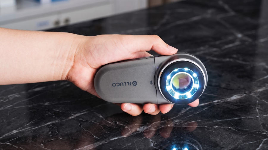
Good technique deserves good optics. The ILLUCO dermatoscope range is built around clarity, comfort and imaging.
· IDS 9100. The flagship of the range, with high optical magnification, cross and parallel polarisation, a UV mode and smartphone imaging, designed for confident, detailed assessment.
· IDS 1100 and IDS 1100C. Polarised dermatoscopes with a wide field of view and strong white LED illumination, the IDS 1100C adding cordless charging.
· IDS 3100 Wood lamp. A dedicated UV tool for pigmentary and infective assessment.
You can compare the full range on the dermatoscopes overview page.
Bringing it together in your practice
The pattern is consistent across every technique. Pick a systematic method, learn it properly, and use equipment that shows you what you need to see. Whether you lean on Chaos and Clues for speed, the two-step algorithm as a structured default, or TADA while you build confidence, the fundamentals hold. Examine deliberately, document well, and keep a clear threshold for biopsy or referral.
If you would like help choosing the right dermatoscope for the way you work, the ILLUCO team can talk you through the range. Book a consultation to get started.
This article is general information for health professionals and is not a substitute for accredited dermoscopy training or individual clinical judgement.


