Australia has one of the highest rates of skin cancer in the world. For GPs, dermatologists, and skin cancer clinicians working on the front line of detection, the quality of the tools in their hands matters enormously, and few factors shape diagnostic quality more directly than the illumination system inside their dermatoscope.
Most conversations about dermoscopy focus on magnification. Lens quality, optical clarity, field of view, these are the specifications that tend to dominate purchasing decisions. But light is where diagnosis actually begins. Without the right illumination, even a superior optical system will miss what matters.
This article explains how modern dermatoscope lighting works, why different light modes serve different clinical purposes, and what UV illumination at 365 nm adds to the equation.
How Light Interacts With Skin
Skin is not a flat surface. Lesions have depth, layering, and irregular texture that respond differently depending on the type of light directed at them. The angle, wavelength, and intensity of illumination each influence what structures become visible, and which ones stay hidden.
This is why contemporary clinical dermatoscopes don't rely on a single light source. They offer multiple illumination modes, each designed to reveal a different layer of diagnostic information.
Polarised Illumination: Cutting Through Surface Glare
Most modern dermatoscopes, including the ILLUCO IDS 9100, offer both cross-polarised and parallel-polarised illumination, switchable during examination.
Cross-polarised light filters out surface reflection, allowing the clinician to see deeper into the dermis without the need for contact fluid. This is particularly useful for visualising vascular structures, regression features, and deeper pigmentation in a fast, comfortable way for the patient.
Parallel-polarised light, used with a liquid interface, emphasises surface structures and the epidermal layers. Subtle features like milia-like cysts and superficial pigment networks become more defined under this mode.
In practice, experienced dermoscopists will often assess a lesion under both modes. Each can reveal features the other obscures, and the clinical picture that emerges from comparing the two is considerably richer than either alone.
UV Illumination at 365 nm: What It Adds
Beyond standard polarised light, UV illumination at 365 nm opens a genuinely different diagnostic channel. At this wavelength, sitting within the UV-A band, certain biological structures, pigments, and microorganisms fluoresce. They absorb the UV light and re-emit it as visible colour, revealing subsurface detail that remains invisible under white or polarised light, non-invasively and in real time.
Pigmentation and sun damage
UV light makes variations in melanin distribution more visually distinct. For Australian clinicians managing patients with significant lifetime UV exposure, this is a practical advantage. Early sun damage, subtle hypopigmentation from previous lesions, and the irregular pigment spread of melasma or vitiligo all become easier to assess and document.
Fungal and bacterial infections
Several common pathogens fluoresce predictably at 365 nm:
- Corynebacterium minutissimum, the organism behind erythrasma, produces a distinctive coral-red glow
- Pseudomonas aeruginosa fluoresces green
- Malassezia species, responsible for pityriasis versicolor, produce a characteristic yellow-green to copper-orange fluorescence, attributed to pityrialactone, a tryptophan-derived metabolite
For a GP or skin clinician seeing a presentation that could fit more than one diagnosis, these visual signatures provide rapid, non-invasive confirmation without waiting on pathology.
Acne assessment
In acne-prone skin, UV examination reveals orange-red fluorescence from porphyrins produced by Cutibacterium acnes. This gives practitioners a more objective way to assess inflammatory activity and track treatment response over time, moving beyond patient self-report to something visible and comparable across visits.
Lesion border definition
UV illumination also enhances contrast at lesion edges and within pigment networks. For early melanoma detection, this added structural definition serves as a useful complement to standard dermoscopic assessment.
Why 365 nm Specifically
The clinical value of UV illumination depends on getting the wavelength right. Shorter UV wavelengths, below approximately 315 nm, in the UV-B band, carry tissue damage risk and aren't appropriate for routine clinical contact. Longer wavelengths, above 400 nm, cross into visible violet light and lose the diagnostic contrast that makes fluorescence clinically useful.
At 365 nm, neither problem applies. The wavelength maximises fluorescence visibility for the chromophores and porphyrins most relevant to skin assessment, produces minimal glare, and penetrates safely without epidermal harm. It has become the accepted standard for clinical UV examination, and the standard wavelength used in modern Wood's lamps, because it balances depth, safety, and diagnostic clarity.
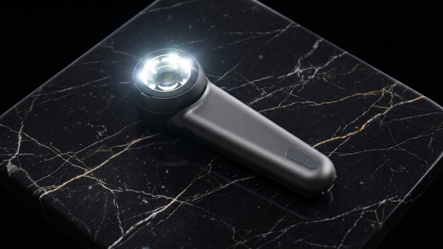
The ILLUCO IDS 9100: All Three Modes in One Device
The ILLUCO IDS 9100 integrates three illumination modes, cross-polarised, parallel-polarised, and UV at 365 nm, into a single handheld device, with four brightness levels per mode for twelve total light settings, giving clinicians precise control over examination conditions.
At 12× optical magnification with high colour accuracy, the IDS 9100 is built for detailed assessment across the full range of skin presentations common in Australian practice. Its full metal housing, rechargeable lithium-ion battery, and infection-control film interface make it equally suited to a busy GP clinic or a specialist dermatology setting.
For clinicians who want to examine the same lesion across multiple illumination modes in a single consultation, and capture accurate images for monitoring or referral, the IDS 9100 is designed to support exactly that workflow.
A Different Way of Seeing
Good dermoscopy has always been about seeing more than the naked eye can. UV illumination at 365 nm extends that principle into territory that conventional light simply cannot reach, revealing pigment disorders, flagging infections, tracking inflammatory activity, and sharpening the boundaries of suspicious lesions.
For Australian clinicians operating in one of the world's highest-risk environments for skin cancer, that extra layer of visibility isn't a luxury. It's a clinical advantage worth having in the room.

Leave a comment
This site is protected by hCaptcha and the hCaptcha Privacy Policy and Terms of Service apply.