


Common Dermatoscopy Mistakes: ILLUCO Corrective Guide
From skipping immersion fluid to pressing too hard, common dermatoscopy mistakes cost diagnostic accuracy. Learn the fixes in ILLUCO's corrective guide.
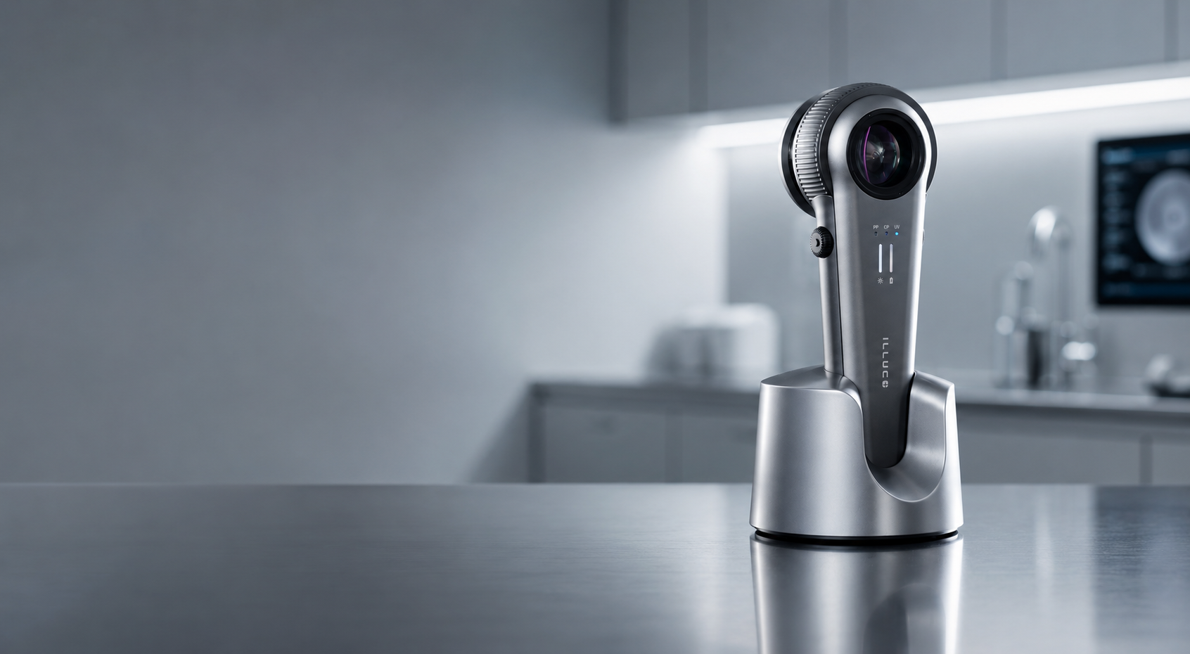
Dermatoscopy has become a core part of skin examination in Australian practice, sharpening diagnostic accuracy for both melanocytic and non-melanocytic lesions. The catch is simple: a dermatoscope is only as good as the technique behind it. Most diagnostic errors fall into two groups, procedural or technical errors made at the device, and knowledge-based mistakes that come down to experience and pattern recognition. This guide from ILLUCO Australia walks through the dermatoscopy mistakes we see most often, and the corrections that fix them.
1. Relying on a single light mode
Polarised and non-polarised light do not reveal the same thing. Polarised dermoscopy is better for dermal vessels, pink and red colours, variable pigmentation and shiny white structures, and it lifts sensitivity for amelanotic or structure-poor melanoma and basal cell carcinoma. Non-polarised dermoscopy is better for superficial features such as milia-like cysts, comedo-like openings and blue-white veil. A seborrhoeic keratosis can look alarming under polarised light yet reassuring under non-polarised light once its milia-like cysts come into view, so leaning on one mode alone invites false positives.
The fix: use both modes and treat them as complementary, not interchangeable. A hybrid dermatoscope that toggles between polarised and non-polarised gives you the full picture. ILLUCO's dermatoscope range covers polarised and contact options, and the IDS 9100 adds cross-polarised imaging.
2. Skipping the immersion fluid, or trapping air bubbles
In contact mode, non-polarised dermoscopy always needs skin contact and a fluid interface. Skip it, or trap air bubbles under the glass plate, and light scatters at the skin-air interface so that structures below the stratum corneum disappear.
The fix: apply enough immersion fluid, whether alcohol gel, ultrasound gel, mineral oil or water, to clear the bubbles and match the refractive index across the glass, fluid and skin. Remember that hybrid devices still need a liquid interface, otherwise only the polarised structures will show.
3. Pressing too hard and blanching the vessels
Cutaneous blood vessels blanch under even slight pressure, so pushing a contact dermatoscope firmly into the skin can wipe out the vascular patterns you are trying to read. That matters because in amelanotic and hypomelanotic lesions, vessel morphology is often the only clue to the diagnosis.
The fix: use minimal pressure, switch to ultrasound gel which cushions the lens, or move to a non-contact polarised view to avoid compressing the lesion at all.
4. Treating dermoscopy as a standalone test
Dermoscopy is part of the clinical examination, not a substitute for it, and errors creep in when it is used in isolation from history and the naked-eye view.
The fix: read each lesion alongside the patient's history and a full skin examination, compare it against the patient's own naevi using the 'ugly duckling' approach, and run a structured method such as the two-step algorithm rather than relying on a gut call.
5. Mistaking artefacts for real structures
Hair, gel residue, surface scale and marker ink can all mimic or obscure genuine dermoscopic structures, and air bubbles in particular back-scatter light and hide what sits beneath.
The fix: clean and prepare the lesion, clear any bubbles, and re-image before you commit to an interpretation.
6. Inconsistent imaging and documentation
Sharp dermoscopic images carry teaching and medicolegal value, and they are the baseline you need to monitor a lesion over time. Blurry or poorly framed captures undercut all of it.
The fix: capture well-framed, in-focus and labelled images, keep a baseline for sequential monitoring, and use a camera or smartphone-enabled device such as the IDS 9100 to make consistent capture part of the routine.
7. Neglecting device hygiene
Dermatoscopes are handled constantly and pressed against skin, and studies have recovered skin flora and Staphylococcus aureus from them, which makes the device a potential source of cross-infection.
The fix: disinfect the contact plate between patients, where 70% isopropyl alcohol is effective, and consider disposable lens covers for higher-risk settings.
8. Under-training and over-relying on one algorithm
Many dermoscopy errors are knowledge-based rather than mechanical, down to limited familiarity and experience, and over-relying on a single rule will miss atypical presentations.
The fix: keep building pattern recognition through ongoing training and CPD, and learn more than one framework so you are not boxed in by a single checklist.
The bottom line
Most dermatoscopy mistakes are not about the eye, they are about setup: the wrong light mode, no immersion fluid, too much pressure or a dirty lens. Correct the technique and the structures speak for themselves. Explore ILLUCO's dermatoscope range to find a device that suits the way you work, or contact the ILLUCO Australia team for help choosing the right setup.


